评估髋臼方向的一种新放射学参数
2019-01-02 文章来源:骨科在线 点击量:1219 我要说
来源:304关节学术
译者:程徽
整理:骨科在线
日本滨松医科大学的Hiroshi Koyama等人发表于《Clinical Orthopaedics & Related》杂志的一项研究显示,利用骨盆正位平片评估髋臼方向时,一种新的定量指标p/a比值是简单可靠的定性指标。
目前定性评估髋臼后倾的方法有多种,但仍缺少定量评估髋臼角度的方法。定量评估将有利于科研,也可能会用于临床协助诊断及治疗髋关节疾病。因此本研究的作者希望找到对髋臼方向采用一种新的定量指标(p/a比),通过测量出平均p/a值,与髋臼后倾放射学征象对比,最终评估其与髋臼解剖方向的关系。

图1A-B(A)p/a 比值由p(髋臼关节面至后壁的距离)除以a(髋臼关节面至前壁的距离)得出,p、a均在根据泪滴低点至髋臼外侧缘连线的垂直平分线上。(B)如上述垂直平分线与髋臼关节面交点在卵圆窝,则是关节面弧形延长线与上述垂直平分线交点作为p、a的近端参考点。

图2A-D(A)正常髋关节。(B)当髋臼前壁轮廓在髋臼后壁的外侧时,为COS阳性。(C)当髋臼后壁边缘在股骨头中心内侧时,PWS为阳性。(D)PRISS征关注坐骨棘是否突入骨盆内,突入则为髋臼后倾。

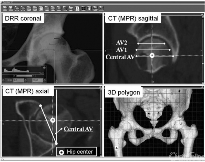
图3:Zed髋关节软件能重建3-D模型,并显示多平面重建(MPR)、数字X线片(DRR)影像,以替代普通平片。在3个垂直于TTP的横截面测量髋臼前倾角:中心前倾角((AV), AV1, AV2),使用DRR,在TPP代表的冠状面上测量p/a比值。
研究人员对骨盆正位平片,测量p(髋臼关节面至后壁的距离)、a(髋臼关节面至前壁的距离),计算p/a值, p、a均在根据泪滴低点至髋臼外侧缘连线的垂直平分线上。他们共 对185例怀疑股骨头坏死的患者进行了测量,并与髋臼后倾的定量指标进行对比。再使用62例无关节炎患者CT,测量其股骨头中部平面处的解剖前倾角,并与p/a比值对比。

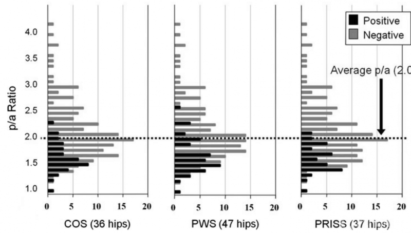
图4:示后倾征分布率,后倾征阳性的髋关节p/a比值很少超过2.05(p<0.001)(185例髋的平均值)。但对于p/a比值小于2.05的髋关节,其后倾征阳性及阴性混杂。

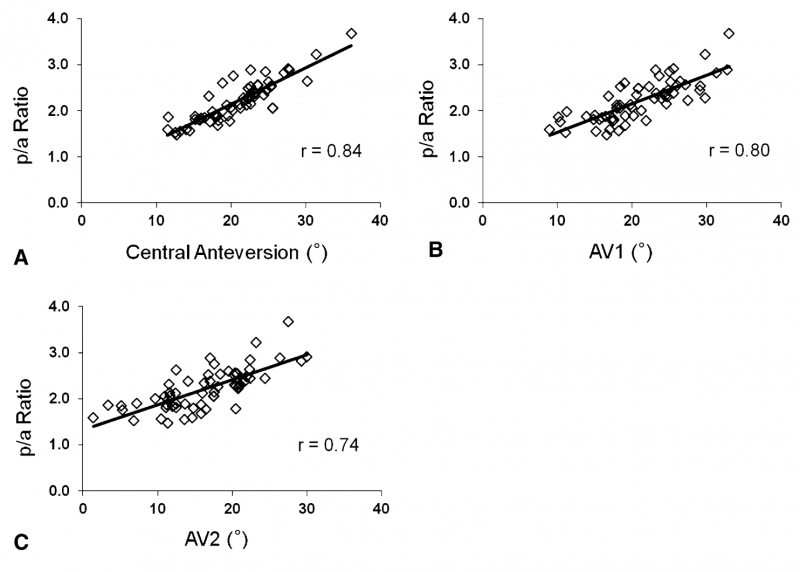
图5A-C:p/a比值与(A) 中心前倾角AV, (B)AV1, (C) AV2的关系。p/a比值与此3个前倾角值均相关(中心前倾角AV: r = 0.84, p<0.001; AV1: r = 0.80, p<0.001; AV2: r = 0.74, p <0.001)。回归分析显示p/a比值与中心前倾角AV存在以下关系:中心前倾角AV=9.6Xp/a –0.3°。
结果显示,185例髋平均 p/a值为2.05,多数大于2.05的患者后倾指标阴性。解剖前倾角与p/a 比值具有相关性(r = 0.84)。
原文摘要:
New Radiographic Index for Evaluating Acetabular Version
Background:
Several qualitative radiographic signs have been describedto assess acetabular retroversion. However, quantitative assessment ofacetabular version would be useful for more rigorous research purposes andperhaps to diagnose and treat hip disorders.
Questions/Purposes:
We developed a new quantitative index for acetabularversion (p/a ratio). We determined the average p/a, compared it with previousradiographic signs for acetabular retroversion, and evaluated its relationship withanatomic acetabular version.
Methods:
We calculated the p/a ratio bymeasuring p (distance from acetabular articular surface to posterior wall) anda (distance from acetabular articular surface to anterior wall) on plain hip AP radiographs and dividing p bya. P and a were assessed on the perpendicular bisector of the line between theteardrop and the lateral edge of the acetabulum. Using 185 hip radiographs frompatients with suspected idiopathic osteonecrosis, we measured p/a and comparedit with previous qualitative signs for acetabular retroversion. Using 62 hip CTimages from patients with no osteoarthritis, we measured the anatomicanteversion at the height of the central femoral head and investigated its relationshipwith p/a.
Results:
The average p/a was 2.05 in 185 hips,and most patients with a p/a greater than 2.05 had a negative qualitativeretroversion sign. A correlation was observed between central anteversion and p/a(r = 0.84).
Conclusions:
We believe this ratio can be considered a simplequantitative parameter to assess acetabular version using plain AP radiographs.
文献出处:
New radiographic index for evaluating acetabular version.Koyama H, Hoshino H, Suzuki D, Nishikino S, Matsuyama Y.ClinOrthop Relat Res. 2013 May;471(5):1632-8. doi: 10.1007/s11999-012-2760-2.


 京公网安备11010502051256号